I still remember the sequence that broke a week of work: five clean injections of mobile A, then a 12-minute gradient, and a ghost peak at RT 7.82 min with an area RSD of 31% across four runs. The peak was not in the blank prepared that morning. It appeared only after the autosampler had been running for six hours. That pattern, an intermittent peak that grows with run time rather than concentration, is the signature of extractables entering the flow path from a consumable. In analytical testing, ghost peaks and contamination are rarely a mystery of chemistry. They are a materials problem with a temperature and a contact time attached. This article is about which compounds bleed from which materials, at which temperatures, and how to prove the source with blank and placebo injections instead of guessing.
A ghost peak is a signal with a retention time and an area, and both are reproducible enough to be measured. That is the key. If the peak is reproducible, it has a source with a fixed geometry and a fixed thermal history. The source is a vial, a septum, a filter, a cap liner, a length of tubing, a column, or a piece of the instrument’s own plumbing. The compound is whatever that material releases under the conditions you impose on it.
I stopped treating contamination as an analytical chemistry problem years ago. It is a materials compatibility problem, and the variables are contact area, contact time, temperature, and solvent strength. Change any one and the bleed rate changes. That is why ghost peaks so often appear late in a run, late in a sequence, or only in the samples that sat in the autosampler tray over a weekend. The chemistry did not change. The exposure did.
The practical consequence is that you can isolate an extractables source without a mass spectrometer, using only blanks and placebos run under controlled conditions. You do need to know what each material class releases, because the retention time and the ion pattern tell you where to look. Start with the consumables, because that is where most of the mass lives. If you are setting up a full analytical testing program and want the consumable side specified correctly from the start, the consumable selection guidance at analytical testing at labanalyticaltesting.com covers the baseline.
What bleeds from what, and at which temperature
The table below is the one I keep taped inside a lab cabinet door. It is not exhaustive, but it covers the materials that cause most ghost peaks in HPLC and GC work. The temperature column is the point at which the material starts to release compounds at a rate that shows up as a peak at typical detector sensitivity (UV at 210 nm, or MS full scan at 106 gain).
| Material | Compounds released | Onset temperature | Typical retention behavior |
|---|---|---|---|
| Silicone (PDMS) septum, general purpose | Cyclosiloxanes (D4, D5, D6), silanols | From 40 °C; strong above 200 °C | Broad, late-eluting humps in GC; silanol tailing in HPLC |
| Silicone/PTFE layered septum | PTFE fluorocarbons, low siloxane | PTFE stable to 260 °C; silicone layer still bleeds | Reduced but not eliminated siloxane |
| Natural rubber / butyl cap liner | Antioxidants, plasticizers, sulfur compounds | From 60 °C | Early sulfur peaks in GC; UV-active species at 210–254 nm |
| Polypropylene (vial, filter housing) | Oligomers, erucamide slip agent, Irgafos-type antioxidants | From 80 °C; worse in strong solvent | Peaks at 5–15 min; m/z 338, 410, 422 common in MS |
| Polyethylene (tubing, caps) | Oligomers, erucamide, BHT | From 70 °C | Similar to PP; BHT at m/z 220 |
| PTFE (tubing, frits, liners) | Very low; trace fluorocarbons at extreme heat | Stable to 260 °C | Minimal bleed; preferred for hot or aggressive work |
| PEEK (fittings, tubing) | Trace oligomers, residual solvent | Stable to 250 °C | Minimal; watch for solvent swelling in DMSO/DMF |
| Cellulose acetate / nylon filter | Membrane extractables, wetting agents | From ambient in aqueous | Early peaks; nylon binds and releases basic analytes |
| Column phase (C18, endcapped) | Silanols, residual silane, phase bleed | From 40 °C; worse above 60 °C | Late humps; gradient baseline rise |
| Septum coring debris | Particulate, then adsorbed analyte | Any | Spikes, split peaks, pressure noise |
Two things stand out. First, silicone is the dominant source in GC and in any heated HPLC autosampler. Cyclosiloxanes are volatile, they are everywhere, and they elute in a characteristic cluster. Second, polypropylene is the dominant source in HPLC, because it is the material of the vial, the filter housing, and half the tubing in the sample path. The erucamide slip agent used in PP molding shows up as a series of peaks at m/z 338 and 410, and it will ruin a low-level MS method if the vials are not specified for LC-MS.
The temperature dependence is steeper than most people assume
Extractables release roughly follows an Arrhenius relationship, which means a 10 °C rise in autosampler tray temperature can double the bleed rate. I have seen a method pass all summer and fail in a cold room, and I have seen the reverse when a tray heater was left on. If your ghost peak appears only in summer, look at the tray temperature before you look at the column. Set the tray to 4–10 °C for labile analytes, and accept that a warmer tray will bleed more from every plastic part in the path.
Isolating the source with blank and placebo injections
You do not need a mass spectrometer to find the source, though it helps. You need a sequence that varies one thing at a time and a definition of a blank that is honest. A blank is not “mobile phase in a vial.” A blank is mobile phase drawn through the same needle, the same loop, the same tubing, and the same vial type as your samples, injected on the same method. If your blank uses a different vial or a different cap, you have not tested the sample path. You have tested the mobile phase.
The four-blank sequence
Run these four in order, and compare the peak at the suspect retention time:
- Blank A, instrument only: no injection, just the gradient. If the peak appears, the source is the column, the mobile phase, or the detector flow cell. Not the sample path.
- Blank B, fresh mobile phase in a new LC-MS grade vial: if the peak appears only here, the source is the vial, cap, or septum.
- Blank C, mobile phase drawn through the full autosampler path (needle, loop, tubing) but from a certified clean vial: if the peak appears here and not in B, the source is the instrument plumbing or a contaminated needle wash.
- Placebo, the sample matrix without analyte, processed exactly as the sample: if the peak appears only here, the source is a filter, a SPE cartridge, a digestion vessel, or a reagent.
The placebo is the one people skip, and it is the one that catches filter and digestion-vessel contamination. A placebo run through the same 0.22 µm filter as the samples will show the membrane extractables at the same retention time as the ghost peak. If it does, the filter is the source, and the fix is a different membrane or a pre-rinse.
Confirming the source with a controlled spike
Once you have a suspect, confirm it with a spike. Take a clean vial, add a known volume of the suspect material (a 1 cm length of tubing, a punched septum disc, a piece of filter membrane), and let it sit in mobile phase at the method temperature for the method’s run time. Inject. If the ghost peak appears at the same retention time and with a comparable area, you have your source. This is the extractables test that most labs never run, and it takes an afternoon.
Field case one: the erucamide peak that only appeared after lunch
Symptom: A contract lab running a USP method for a small-molecule API saw a peak at RT 6.4 min that appeared in about one in five injections, always in the afternoon, never in the morning. Area varied from 0.02 to 0.15 mAU·s. The peak was not in the morning blank.
Measurement: The lab ran the four-blank sequence. Blank A (instrument only) was clean. Blank B (fresh mobile phase in an LC-MS grade vial) was clean. Blank C (mobile phase drawn through the autosampler path) showed the peak. The placebo, filtered through the lab’s standard 0.22 µm PP filter, also showed it. The retention time matched an erucamide standard at m/z 338 in the MS confirmation run.
Root cause: The autosampler needle wash vial was a general-purpose PP vial, and the wash solvent was 50:50 water:acetonitrile. Over a morning of injections the wash vial sat at room temperature, and the PP released erucamide into the wash. The needle carried a film of it into the next injection. Afternoon runs were worse because the wash had been sitting longer.
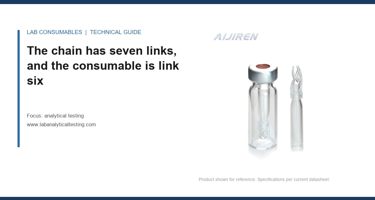
Fix: Switch the wash vial to a certified low-extractable PP or glass vial, and change the wash every four hours. The peak disappeared within one sequence. The lab now specifies LC-MS grade vials for the wash, not just for the samples. The vial specification details are on the chromatography vial page, and the difference between general-purpose and LC-MS grade is the molding resin, not the label.
Field case two: the siloxane hump that scaled with tray temperature
Symptom: A GC-MS method for residual solvents showed a broad hump at 18–22 min that was absent in the solvent blank but present in every sample. The hump area correlated with how long the samples sat in the tray, not with analyte concentration. The lab had already replaced the column and the liner.
Measurement: The lab ran a septum extractables test: a single septum was placed in a sealed headspace vial and heated to 80 °C for 30 minutes. The resulting chromatogram showed the same hump, with the D4, D5, and D6 cyclosiloxane pattern at m/z 281, 355, and 429. The tray heater had been set to 45 °C to keep samples warm, and the septa were general-purpose silicone, not the PTFE-faced type.
Root cause: The tray heater was raising the septum temperature above the point where silicone begins to release cyclosiloxanes at a detectable rate. The samples that sat longest bled the most. The column and liner were innocent.
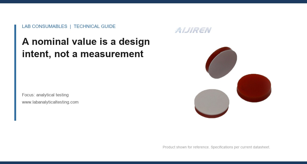
Fix: Switch to PTFE-faced septa (the PTFE face toward the sample), turn the tray heater down to 25 °C, and cap the samples immediately after preparation. The hump dropped below the reporting limit. The lab now specifies PTFE-faced septa for any method with a tray temperature above 30 °C, and it checks the septum lot against a reference standard before use. The consumable options are catalogued at HPLC vial factory, including the PTFE-faced and pre-slit variants.
The consumable specifications that prevent most ghost peaks
Most ghost peaks I have traced back to a consumable were preventable at the point of purchase. The specification is not just “clean.” It is a set of numbers that a supplier can meet and a lab can verify. Here is the checklist I use when qualifying a consumable for a method that runs below 1 ppm or uses MS detection.
| Parameter | Target for trace/MS work | Why it matters |
|---|---|---|
| Vial resin | LC-MS grade PP or borosilicate glass, Class A | General PP releases erucamide and oligomers |
| Septum type | PTFE-faced silicone or silicone/PTFE layered | Bare silicone bleeds cyclosiloxanes above 40 °C |
| Septum durometer | Shore A 45–55 | Softer cores; harder leaks. 45–55 balances both |
| Puncture count | Replace after 1–3 punctures in GC; 5–10 in HPLC | Coring debris and leak paths grow with each puncture |
| Cap torque | 8–12 in·oz for 9 mm screw caps; 4–6 in·oz for 11 mm crimp | Over-torque deforms the septum and opens a bleed path |
| Vial cleanliness | Certified < 1 ppb extractables by LC-MS | The certificate is the only way to know the lot |
| Filter membrane | PTFE or PVDF, pre-rinsed, low extractables | Cellulose acetate and nylon release wetting agents |
| Tubing | PTFE or PEEK, 0.005–0.010 in ID | PE tubing bleeds oligomers; larger ID adds dead volume |
The puncture count is the one people ignore. A septum rated for 10 punctures is not a suggestion. I have seen a lab run 40 injections through a single GC septum and wonder why the baseline drifted and the peak areas fell. Each puncture opens a channel, and the channel is a leak path and a bleed path. Replace the septum on a schedule tied to injection count, not to the calendar. For GC, the schedule is often every 50–100 injections for a PTFE-faced septum, and every 25–50 for a general-purpose one.
The cap torque matters more than most labs realize. A 9 mm screw cap torqued to 15 in·oz compresses the septum into the bore and can extrude it, which produces a bleed path and a coring risk. A cap torqued to 4 in·oz is loose enough to leak. The 8–12 in·oz range is the one that seals without deforming, and a torque screwdriver is a $80 tool that pays for itself in one avoided investigation. The dimensional standards for these caps are covered in the chromatography products catalog, including the GPI 9 mm and 11 mm thread specifications.
Where the instrument itself contributes
The consumable is the usual suspect, but the instrument has its own extractables sources. The column is one. A C18 column that has been heated above its rated temperature will bleed phase and silanols, and the bleed shows up as a rising baseline and late humps in gradient runs. Check the column’s temperature rating, and do not run it above 60 °C unless it is rated for it. The detector flow cell is another. A flow cell with a scratched window or a trapped bubble will show a peak that moves with flow rate, not with retention time.
The mobile phase is the third. Water from a system that has not been serviced in a year can carry bacterial byproducts and plasticizer from the tubing. I have traced a ghost peak at 210 nm to a length of PE tubing between the water polisher and the HPLC. The fix was to replace it with PTFE. The general guidance from the column and consumable manufacturers is worth reading before you blame the instrument: Agilent’s chromatography consumables and columns documentation lists the bleed profiles for their phases, and Thermo Fisher’s chromatography and sample handling resources cover the sample path materials. For method-level context on how extractables are handled in regulated work, the U.S. EPA SW-846 test methods are a useful reference for holding times and container requirements, and Restek’s technical resources have practical notes on septum and liner selection that match what I have seen in the field.
A troubleshooting checklist you can run in one shift
When a ghost peak appears, work through this list in order. Each step narrows the source by one material class.
- Step 1: Confirm the peak is reproducible. Run the same blank three times. If the area RSD is above 20%, the source is likely a variable exposure (tray time, wash age) rather than a fixed material.
- Step 2: Run blank A (instrument only). If the peak is present, the source is the column, mobile phase, or flow cell. Stop here and check the column bleed and the water system.
- Step 3: Run blank B (fresh mobile phase in a certified vial). If the peak appears, the source is the vial, cap, or septum.
- Step 4: Run blank C (full autosampler path). If the peak appears here but not in B, the source is the needle, loop, wash, or tubing.
- Step 5: Run the placebo. If the peak appears only in the placebo, the source is a filter, SPE cartridge, digestion vessel, or reagent.
- Step 6: Spike test the suspect. Soak a piece of the suspect material in mobile phase at method temperature for the run time and inject. Match the retention time and area.
- Step 7: Fix and verify. Change one consumable, rerun the four-blank sequence, and confirm the peak is below the reporting limit before releasing the method.
The checklist is deliberately ordered from the cheapest test to the most expensive. Steps 1 through 5 cost a few hours of instrument time. Step 6 costs an afternoon. Replacing a column or an instrument module without running steps 1 through 5 is the most common way labs waste a week.
The decision rule for consumable qualification
If a method reports below 1 ppm, uses MS detection, or runs a gradient, qualify every consumable in the sample path against an extractables specification before the method is validated. That means LC-MS grade vials, PTFE-faced septa, PTFE or PEEK tubing, and pre-rinsed filters, each with a certificate and each verified by a blank injection at the method’s temperature and run time. The specification is the control; the blank is the proof. When a ghost peak appears, run the four-blank sequence and the placebo before you touch the instrument, because the source is almost always a material with a temperature rating you exceeded or a puncture count you ignored.
References & further reading
Related resources from our lab consumables network:
- Analytical Testing at labanalyticaltesting.com
- Chromatography Vial at chromvials.com
- Hplc Vial at hplcvialfactory.com
- Chromatography Products at chromatographyforsale.com
External references and standards: